


 FDA staining of batch microalgal cultures at different growth stages
Fluorescein diacetate (FDA) hydrolysis assays can be used to measure enzyme activity produced by microbes in a sample. "Fluorescein and fluorescein derivatives are widely used because they can be excited at the popular 488-nm line of anargon laser, because they have high extinction coefficients and high quantum yields (although pH dependent), and because they may be easily loaded into intact, esterase-containing (viable) cells by incubation with the respective diacetyl esters. The fluorescence signal of cells loaded with fluorescein orf luorescein derivatives depends largely on the intracellular concentration of the probe and the pH. Basically, this concentration is the result of uptake and hydrolysis of the nonfluorescent precursor and efflux of the fluorescent product. It is generally assumed that nonpolar esterified compounds such as FDA diffuse freely into intact cells, although this has never been verified. However, an active uptake mechanism for FDA in erythrocytes, CHO cells, and HeLa cells has been suggested. After transport into the cells, the prefluorochrome is hydrolyzed into the more polar fluorescent products by intracellular esterases. Apparently, most cells, whether mammalian cells, yeasts cells, or gram-positive or gram-negative bacteria, can hydrolyze FDA or cFDA" (Breeuwer et al, 1995).
Ekaterina Solomonova, a PhD student at the Dept of Algal Ecophysiology (IBSS), and Vladimir Mukhanov, a research scientist at Dept of Plankton (IBSS), stained batch microalgal cultures with FDA to measure the ratio between active (FDA+) and inactive or dead cells (FDA-) over different phases of their growth (Fig. 1).
Ekaterina Solomonova, a PhD student at the Dept of Algal Ecophysiology (IBSS), and Vladimir Mukhanov, a research scientist at Dept of Plankton (IBSS), stained batch microalgal cultures with FDA to measure the ratio between active (FDA+) and inactive or dead cells (FDA-) over different phases of their growth (Fig. 1).
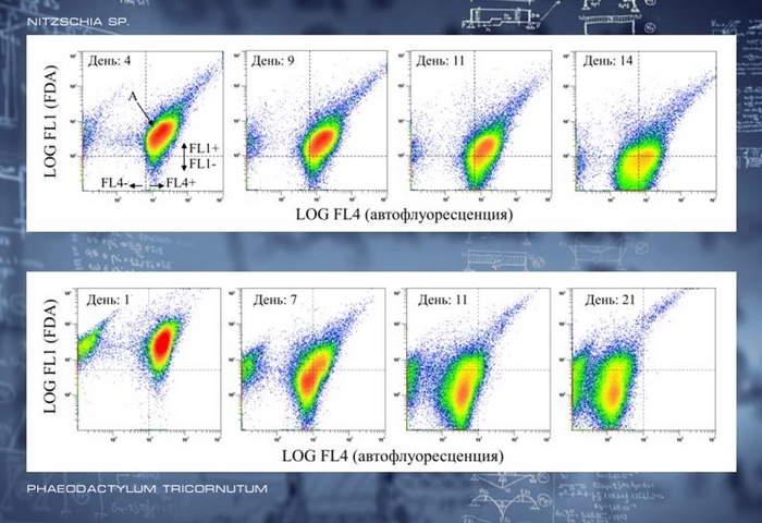
Fig. 1. Decrease in cell enzyme activity (FL1 – FDA fluorescence) and pigment content (FL4 - chlorophyll autofluorescence) in batch Nitzschia sp.(top plots) and Phaeodactylum tricornutum (bottom) cultures. А – claster of the cultured microalgae. Dashed lines differ four cell subpopulations: alive, physiologically active cells (FL1+); non-active and dead cells (FL1); cells with high (FL4+) and low (FL4) pigment content. Age of the cultures in days is presented. Data are provided by Vladimir Mukhanov and Ekaterina Solomonova.
Back to the list of applications
